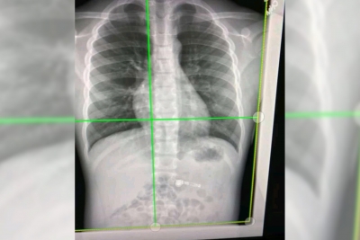
High-energy X-rays show lung vessels altered by Covid-19
By IANS | Published: November 5, 2021 12:45 PM2021-11-05T12:45:03+5:302021-11-05T12:55:38+5:30
London, Nov 5 Using high-energy X-rays, scientists have found how Covid-19 damages even the smallest blood vessels in ...

High-energy X-rays show lung vessels altered by Covid-19
London, Nov 5 Using high-energy X-rays, scientists have found how Covid-19 damages even the smallest blood vessels in human lungs.
Scientists from University College London and the European Synchrotron Research Facility (ESRF) used a new revolutionary imaging technology called Hierarchical Phase-Contrast Tomography (HiP-CT), to scan donated human organs, including lungs from a Covid-19 donor.
Using HiP-CT, the research team saw how severe Covid-19 infection 'shunts' blood between the two separate systems the capillaries which oxygenate the blood and those which feed the lung tissue itself.
Such cross-linking stops the patient's blood from being properly oxygenated, which was previously hypothesised but not proven, said the team in the paper published in the journal Nature Methods.
"
The HiP-CT technique provides the brightest source of X-rays in the world at 100 billion times brighter than a hospital X-ray.
Due to this intense brilliance, researchers can view blood vessels five microns in diametre (a tenth of the diametre of a hair) in an intact human lung, whereas a clinical CT scan only resolves blood vessels that are about 100 times larger, around 1mm in diametre.
The UCL-led team is now planning to use HiP-CT to produce a Human Organ Atlas. This will display six donated control organs: brain, lung, heart, two kidneys and a spleen, and the lung of a patient who died of Covid-19. There will also be a control lung biopsy and a Covid-19 lung biopsy. The Atlas will be available online for surgeons, clinic and the interested public.
The researchers believe that the scale-bridging imaging from whole organ down to cellular level could provide additional insights into many diseases such as cancer or Alzheimer's Disease.
Disclaimer: This post has been auto-published from an agency feed without any modifications to the text and has not been reviewed by an editor
Open in app